This web page is to inform about the vision, engage researchers and potential future users, and encourage discussion and exchange. The state of the project is: basic research project.
We propose a revolutionary new way to guide radiological interventions
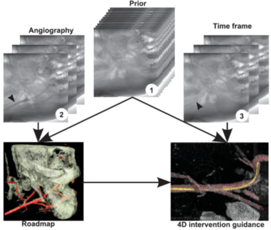
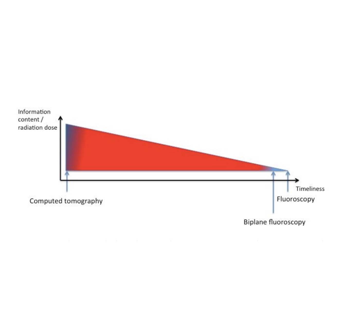
Find Out More4D intervention guidance would provide a superposition-free situational awareness at times, while still providing 2D fluoroscopy as in today`s C-arms (with only one degree of freedom lost).
4D intervention guidance might allow for rethinking how interventions are done. As a result, they can become faster and safer. Additionally, currently, unforeseeable and completely novel approaches can be developed.
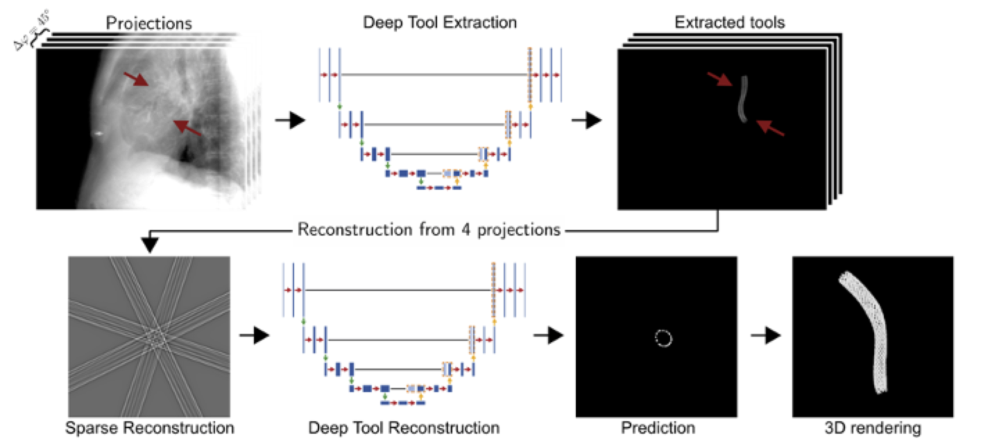
Several challenges need to be addressed in research. For example, there are geometrical challenges, short but high photon flux X-ray flashes are necessary, and fast, high-power reconstruction and machine learning algorithms need to be implemented in real-time.
Acquiring a 4D data set is only the first step. The data also needs to be processed and displayed in a practical and accessible way. Uncertainties in the dataset need to be visualized. (Remark: While 3D goggles are a fancy toy, we don't see a future for them in the CATH lab since a 2D screen can visualize an intervention situation sufficiently good.)
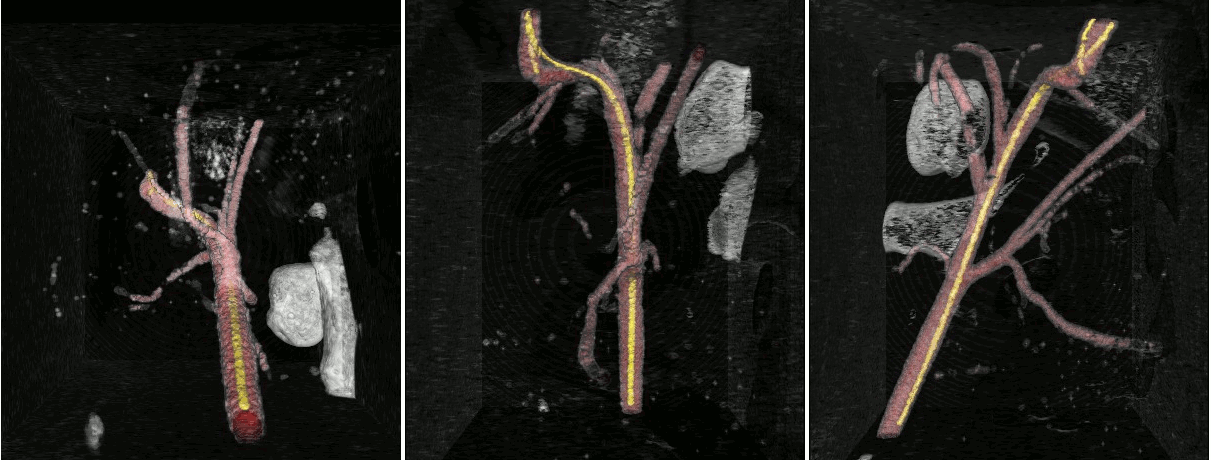
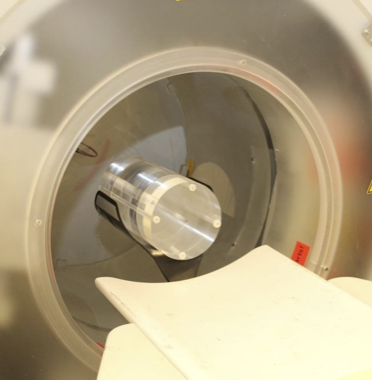
While we have acquired promising results in early simulations, phantom scans, and in-vivo experiments, it is essential to understand that 4D intervention guidance is currently a research vision.
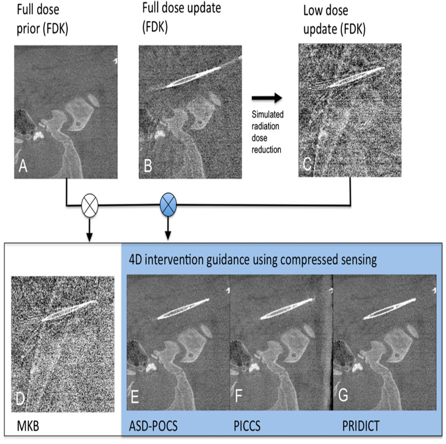
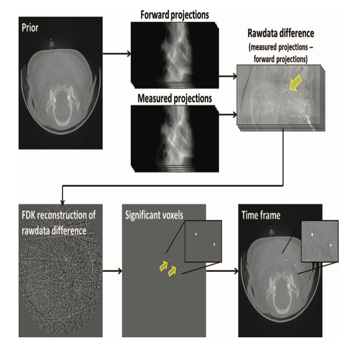
Radiation dose is the first thing that comes up. This is the challenge we are up to - however, on our side are: novel reconstruction algorithms, sparse volume changes during interventions, and potentially shorter interventions. Please see our publications for discussion. We don't do "CT-Fluoroscopy"!
The convergence of novel X-ray sources, new detectors, slim gantries, fast computers, novel reconstruction algorithms, and machine learning will make this vision reality.
While C-arms provide better access to the patient, at first sight, it is just too dangerous for them to "wobble" around in the Cathlab. Furthermore, in real life, their movement is restricted through sterile covers, lines, and infusions, so in the most critical situations, we still don't acquire 3D datasets... when we would need them the most ...
The inventors Prof. Marc Kachelrieß, PD Dr. Sönke Bartling and Dr. Jan Kuntz worked with cone-beam beforehand. On background of the arrival of interventional C-arm systems that are capable of CT-imaging the first exploratory experiments were performed on VCT. Successful DFG proposal. A patent was filed.
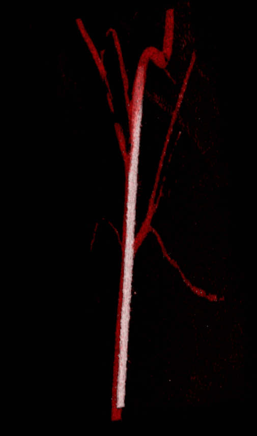
A general description of the concept of 4D intervention guidance was published in European Radiology. Describing the first experimental ex-vivo and in-vivo results compromising an experimental continuously rotating flat-panel Volume-CT setup with simulated sparse sampling.
Published in Medical Physics and Physics in Medicine & Biology
Thanks to the German Neuroradiological society.
An overview publication was published on Arxive.org describing the advantages and disadvantages of 4D intervention guidance and discussing the actual implementation in a cathlab.
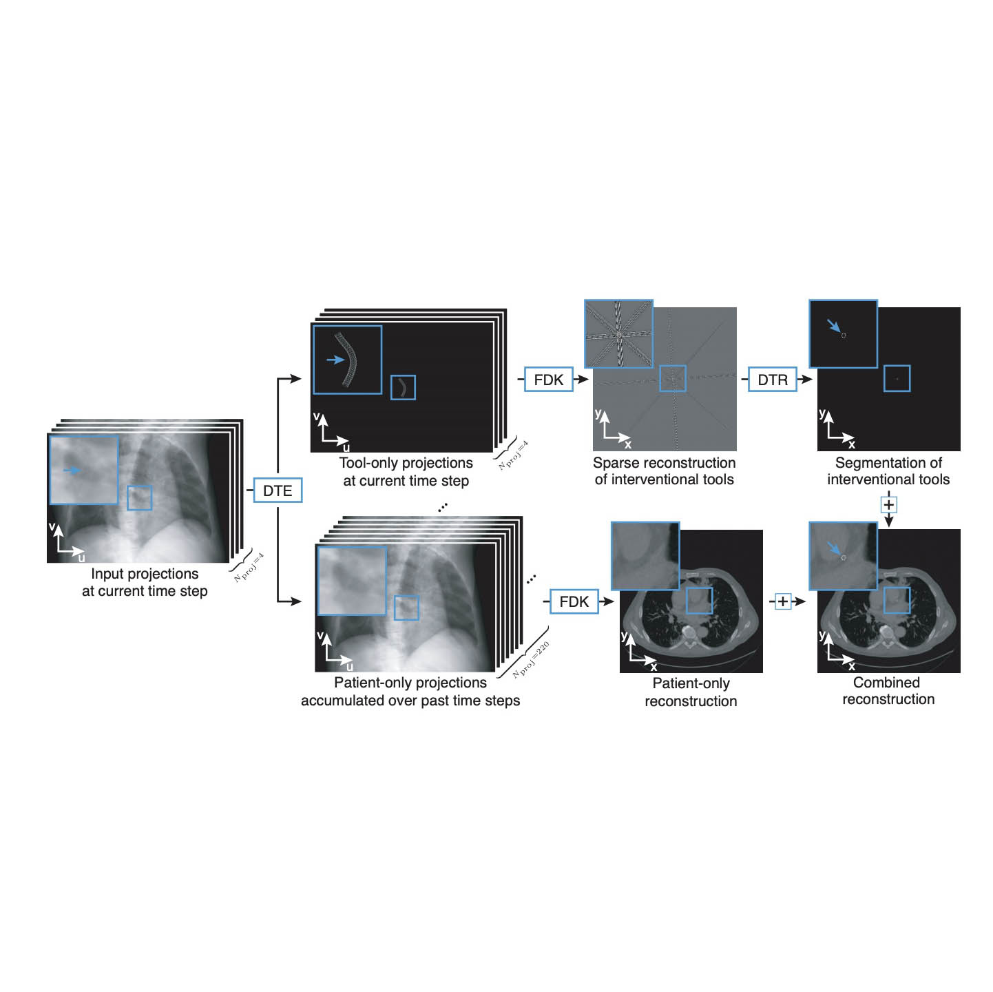
Machine learning can be used to exploit the constraints in interventional tomographic imaging